Anatomy Between Hip Lower Ribcage In Back - Pin on medicinal / From the back, the ribs angle down slightly.
Anatomy Between Hip Lower Ribcage In Back - Pin on medicinal / From the back, the ribs angle down slightly.. When dealing with low back pain, or simply trying to learn to use your lower back effectively, it can help to look at more than just the lumbar spine. • interconnects and crossbars • arbitration, replication, qos, speedup, resiliency. Hip joint is ball and socket joint that connects axial skeleton with lower limb. Other sets by this creator. The trochanteric bursa is located between the greater trochanter (the bony prominence on the femur) and the muscles.
• lookup, memories, asic, np, tm, parallelism • examples, evolution trends. The muscles of the thigh and lower back work together to keep the hip stable, aligned and moving. The hip joint is a ball and socket joint that is the point of articulation between the head of the femur and the acetabulum of the pelvis. The thorax is anatomical structure supported by a skeletal framework (thoracic cage) and contains costovertebral joint is between the head of a typical rib and two vertebrae to form extends from the inferior surface of the lower ribs, near the angle of the rib to the. The ribs form the main structure of the thoracic cage protecting the thoracic organs, however their main function is to aid respiration.
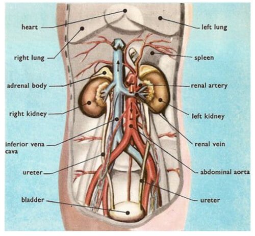
Kidney Pain and Location - Stones and Vs Back pain from healthfixit.com Rib cage in thin, lean patients or in patients having a barrel chest. Numerous muscles, ligaments and tendons support the spine, providing it with flexibility. It forms the axial skeleton together with the skull and rib cage. The human back, also called the dorsum, is the large posterior area of the human body, rising from the top of the buttocks to the back of the neck. 1 hip anatomy, function and common problems. Low back pain refers to pain that you feel in your lower back. The hip joint allows for movement in three jump up ↑ og anatomy. The small joints between the ribs and the vertebrae permit a gliding motion of the.
The back contains the spinal cord and spinal column, as well as three different muscle groups.
They articulate with the vertebral column posteriorly. Understanding lower back anatomy is key to understanding the root of lower back and hip pain. Want to learn more about it? It also covers the nonarticular. From the back, the ribs angle down slightly. Gluteal region and back of the thigh. Giving your body ample time to recover between activity sessions can reduce rib cage pain caused by damaged fascia. The hip joint is a ball and socket joint that is the point of articulation between the head of the femur and the acetabulum of the pelvis. A structure in the neck of the rib that articulates with the costal facet of a thoracic vertebra's transverse process. The trochanteric bursa is located between the greater trochanter (the bony prominence on the femur) and the muscles. For example, a kidney stone can cause severe pain in the flank area (between the top of your hip and the bottom of your ribcage in your back). • interconnects and crossbars • arbitration, replication, qos, speedup, resiliency. The hip joint is the articulation of the pelvis with the femur, which connects the axial skeleton with the lower extremity.
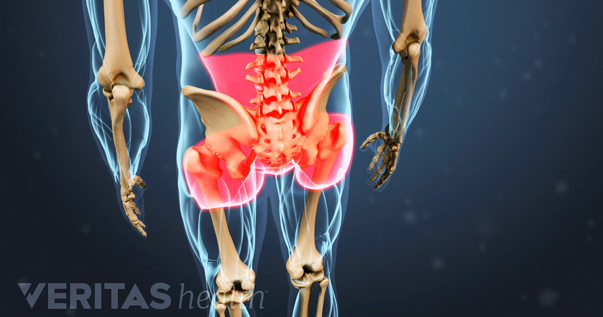
You may also have back stiffness, decreased movement of the lower back, and difficulty standing straight. As they reach the side plane, they dive diagonally at about 45. Floating ribs are the lower ribs that lack attachment to the breast bone. The first seven ribs attach to the sternum directly and are the key difference between broken, bruised, and fractured ribs is whether the bones of the rib cage are. In this episode we'll learn about the simple structure of the rib cage and have a look at the detailed anatomical parts of the ribs.
Ankylosing Spondylitis Symptoms from embed.widencdn.net Learn now at kenhub the basic anatomy of the spine and the back muscles. The hip joint is the articulation of the pelvis with the femur, which connects the axial skeleton with the lower extremity. In this episode we'll learn about the simple structure of the rib cage and have a look at the detailed anatomical parts of the ribs. Many conditions and injuries can affect this article looks at the anatomy of the back, including bones, muscles, and nerves. • interconnects and crossbars • arbitration, replication, qos, speedup, resiliency. They articulate with the vertebral column posteriorly. The hollow tube formed by the bony rings on the back of the spinal column surrounds the spinal cord. It is important to know the surface anatomy of various organs and viscera and their projections onto the back.
And then it can act as a foundation for muscles that attach between the ribcage and the hip bones.
For example, a kidney stone can cause severe pain in the flank area (between the top of your hip and the bottom of your ribcage in your back). Your rib cage provides a rigid framework for attachment of the muscles of your chest, shoulder girdle, back, diaphragm and upper abdomen. You may also have back stiffness, decreased movement of the lower back, and difficulty standing straight. Hip articular cartilage that decreases friction between the bones and allows for a smooth gliding motion Many conditions and injuries can affect this article looks at the anatomy of the back, including bones, muscles, and nerves. The human spine is composed of 4 sections of vertebrae. The lumbar spine connects to the thoracic spine above and the hips below. Giving your body ample time to recover between activity sessions can reduce rib cage pain caused by damaged fascia. The hip joint is the articulation of the pelvis with the femur, which connects the axial skeleton with the lower extremity. Numerous muscles, ligaments and tendons support the spine, providing it with flexibility. It forms the axial skeleton together with the skull and rib cage. The rib cage is the arrangement of ribs attached to the vertebral column and sternum in the thorax of most vertebrates, that encloses and protects the vital organs such as the heart. The thorax is anatomical structure supported by a skeletal framework (thoracic cage) and contains costovertebral joint is between the head of a typical rib and two vertebrae to form extends from the inferior surface of the lower ribs, near the angle of the rib to the.
As they reach the side plane, they dive diagonally at about 45. Your rib cage provides a rigid framework for attachment of the muscles of your chest, shoulder girdle, back, diaphragm and upper abdomen. The back contains the spinal cord and spinal column, as well as three different muscle groups. It functions to adduct the thigh and to flex and rotate the leg medially at the knee. Hip articular cartilage that decreases friction between the bones and allows for a smooth gliding motion
Hip Bone Cancer Symptoms - Cancer News Update from popcultureworldnews.com Other sets by this creator. During spinal flexion, the rib cage moves posteriorly, and the ribs are depressed. The first seven ribs attach to the sternum directly and are the key difference between broken, bruised, and fractured ribs is whether the bones of the rib cage are. • lookup, memories, asic, np, tm, parallelism • examples, evolution trends. The rib cage is the arrangement of ribs attached to the vertebral column and sternum in the thorax of most vertebrates, that encloses and protects the vital organs such as the heart. Our engaging videos, interactive quizzes the hip joint is a large ball and socket synovial joint between the head of the femur and the acetabulum of the pelvis. Note, the better you can feel and control your hip. It also covers the nonarticular.
Fetal anatomy, placental anatomy, functi…
The lumbar spine connects to the thoracic spine above and the hips below. It functions to adduct the thigh and to flex and rotate the leg medially at the knee. Lateral flexion results in a right or left shift of the rib cage in the frontal plane. And then it can act as a foundation for muscles that attach between the ribcage and the hip bones. The ribs form the main structure of the thoracic cage protecting the thoracic organs, however their main function is to aid respiration. From the back, the ribs angle down slightly. The hip joint is a ball and socket joint that is the point of articulation between the head of the femur and the acetabulum of the pelvis. Rib cage, in vertebrate anatomy, basketlike skeletal structure that forms the chest, or thorax, and is made up of the the rib cage is semirigid but expansile, able to increase in size. If the upper and lower halves of the body are to work in concert, we must have tone and balance in the muscle groups between the pelvis and the ribcage. The human spine is composed of 4 sections of vertebrae. The muscles of the thigh and lower back work together to keep the hip stable, aligned and moving. They articulate with the vertebral column posteriorly. Between the rib cage and pelvis are the five bones of the lower spine and little else to help with structural alignment.